Cataracts Explained: Symptoms, Surgery Options, and Recovery Guide

Imagine looking at the world through a dirty window. Colors seem duller, lights leave halps around them, and reading your favorite book requires squinting until your eyes burn. This isn't just aging; it’s likely cataracts, a progressive clouding of the eye's natural lens that blurs vision. You are not alone in this struggle. According to data from the National Eye Institute, approximately 24.4 million Americans aged 40 and older were affected by cataracts as of 2019. It is the leading cause of vision loss globally, but unlike many other eye conditions, it is entirely treatable.
The good news? Cataract surgery is one of the most successful medical procedures performed today. With a patient satisfaction rate of 98% according to a 2021 survey by the American Society of Cataract and Refractive Surgery (ASCRS), millions of people regain their clarity every year. But knowing when to get surgery, what type of lens to choose, and what recovery looks like can be overwhelming. Let’s break down exactly what happens during the process so you can make an informed decision without the anxiety.
Understanding What Causes Cloudy Vision
Your eye has a clear lens behind the iris that focuses light onto the retina at the back of your eye. Think of it like the lens on a camera. When you have cataracts, proteins inside this lens begin to clump together. Over time, these clumps form cloudy spots that block light from passing through clearly. While age is the primary culprit-often starting in your 40s or 50s-it’s not the only factor.
Other contributors include prolonged exposure to ultraviolet sunlight, smoking, diabetes, and previous eye injuries. Some medications, particularly long-term use of corticosteroids, can also accelerate the formation of cataracts. The condition develops slowly, which is why many people don’t realize they have it until daily tasks become frustrating. You might notice that night driving becomes dangerous due to glare from oncoming headlights, or that colors appear faded, like an old photograph losing its vibrancy.
Historically, treating cataracts was risky. In ancient times, surgeons used a technique called 'couching,' where they simply pushed the lens out of place without replacing it. That changed forever in 1949 when Sir Harold Ridley implanted the first intraocular lens (IOL) at St Thomas' Hospital in London. This innovation transformed cataract treatment from a last resort into a routine procedure that restores functional vision.
When Is Surgery Actually Necessary?
A common misconception is that you should wait until the cataract is 'ripe' or fully mature before getting surgery. This is outdated advice. Today, the decision to operate is based on how much the cataract interferes with your quality of life. If you find yourself struggling to read menus, recognize faces, or drive safely, it may be time to talk to your ophthalmologist.
Surgery is typically recommended when:
- Vision impairment affects daily activities: You can no longer perform tasks you enjoy, such as gardening, knitting, or playing sports.
- Night driving becomes unsafe: Glare and halos make it difficult to see road signs or pedestrians.
- Professional demands require sharp vision: Jobs requiring precision, such as piloting or detailed craftsmanship, suffer due to blurred sight.
- Other eye conditions are harder to manage: Dense cataracts can prevent doctors from examining the retina for issues like macular degeneration or diabetic retinopathy.
There is no strict timeline. Some people live with mild cataracts for years using stronger glasses, while others seek surgery sooner to maintain independence. The key is open communication with your eye care provider about your specific needs.
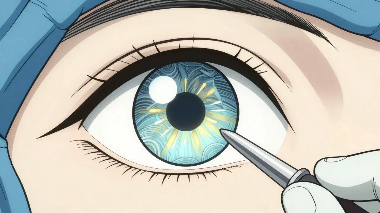
The Procedure: Phacoemulsification and Lens Implants
Modern cataract surgery is remarkably efficient. About 95% of procedures today use a technique called phacoemulsification, a surgical method that uses ultrasound waves to break up the cloudy lens. Here is what happens step-by-step:
- Anesthesia: You receive local anesthesia via drops or an injection to numb the eye. You remain awake but feel no pain.
- Incision: The surgeon makes a tiny incision, usually between 2.2mm and 2.8mm, in the cornea. This small size means stitches are rarely needed.
- Fragmentation: An ultrasonic probe is inserted through the incision. It emits high-frequency sound waves that break the cloudy lens into tiny pieces.
- Removal: These fragments are suctioned out gently.
- IOL Insertion: A foldable artificial lens, known as an intraocular lens (IOL), an artificial lens implanted in the eye to replace the natural lens, is inserted into the empty lens capsule. It unfolds automatically once inside.
The entire process takes about 10 to 20 minutes per eye. Most patients go home the same day. Because the incision is so small, healing is fast, and the risk of infection is significantly lower than in older surgical methods.
Choosing Your Artificial Lens: Monofocal vs. Premium Options
One of the biggest decisions you will face is selecting the type of IOL. This choice impacts your dependence on glasses after surgery. Standard insurance plans, including Medicare, typically cover only basic monofocal lenses. However, premium options offer enhanced visual outcomes for those willing to pay out-of-pocket.
| Lens Type | Focus Range | Glasses Needed After? | Estimated Cost Per Eye (Out-of-Pocket) |
|---|---|---|---|
| Monofocal | Distance only | Yes, for near/intermediate | $0 (covered by insurance) |
| Toric | Distance + Astigmatism correction | Less likely for distance | $2,500 - $4,500 |
| Extended Depth of Focus (EDOF) | Distance + Intermediate | Maybe for close reading | $3,000 - $4,500 |
| Trifocal/Multifocal | Distance, Intermediate, Near | Rarely (81-89% spectacle independence) | $3,500 - $5,000+ |
If you rely heavily on computers or enjoy hobbies like golf or tennis, an EDOF or trifocal lens might be worth the investment. Brands like Alcon’s PanOptix and Johnson & Johnson’s Tecnis Symfony have gained popularity for offering excellent range of vision. However, if you prefer simplicity and don’t mind wearing reading glasses, a standard monofocal lens works perfectly well and carries fewer risks of visual disturbances like halos.

Recovery Timeline: What to Expect Week by Week
Recovery varies from person to person, but there is a general pattern. Immediately after surgery, your eye may feel scratchy, sticky, or slightly uncomfortable. Mild itching is normal for a couple of days. Vision might look blurry or hazy right away, but this improves gradually.
Most people notice significant improvement within 1 to 3 days. By the end of the first week, many return to non-strenuous work. Full stabilization of vision can take anywhere from four weeks to three months. During this time, your brain adapts to the new lens, especially if you had different prescriptions in each eye previously.
To ensure smooth healing, follow these post-operative rules strictly:
- No rubbing: Avoid touching or rubbing your eye for at least two weeks.
- Water safety: Keep water out of your eye when showering or washing your face for the first week.
- Medication adherence: Use prescribed antibiotic drops (like moxifloxacin) for one week and steroid drops (like prednisolone acetate) tapered over four weeks to prevent inflammation.
- Activity restrictions: Avoid heavy lifting, rigorous exercise, and swimming for several weeks.
- Driving: Do not drive until your doctor clears you, usually after a follow-up visit confirms safe vision levels.
If you experience severe pain, sudden vision loss, or increased redness, contact your surgeon immediately. These could signal complications, though they occur in less than 5% of cases.
Potential Complications and Long-Term Outlook
While cataract surgery is incredibly safe-with a 99.5% safety rate according to a 2022 meta-analysis in the British Journal of Ophthalmology-risks do exist. One common issue is posterior capsule opacification (PCO), often called a 'secondary cataract.' This happens when the membrane holding the IOL becomes cloudy months or years later. It affects 20-30% of patients within five years but is easily treated with a quick YAG laser capsulotomy procedure in the office.
Other rare complications include infection, retinal detachment, or persistent inflammation. Patients with pre-existing conditions like glaucoma or diabetes may experience slower healing or incomplete vision restoration. In these cases, additional therapies, such as vision therapy exercises to retrain the brain-eye connection, can help maximize outcomes.
Long-term, the success rate is outstanding. Approximately 95% of patients report significant vision improvement. Many describe the experience as seeing 'HD' for the first time in decades, with vibrant colors and sharper contrast. As technology advances, future lenses may even mimic the natural accommodation of the young eye, further reducing reliance on corrective eyewear.